The heart is the first functional organ in a vertebrate embryo. There are 5 stages to heart development.
- Primum Step 3 Program Schedule
- Primum Step 3 Program Reviews
- Primum Step 3 Program Manual
- Primum Step 3 Program Download
Excerpts from writings about how step 3 of the 12 step program works. STEP 3 assesses whether you can apply medical knowledge and understanding of biomedical and clinical science essential for. Pathway program (at a date. This isn’t required under the VA Home Loan Program, but it’s certainly a good idea — especially for the first-time home buyer. Of all the 12 Steps, Step 5 is probably the hardest for everyone. It's perfectly natural to fear it. Learn why admitting our wrongs is a key to recovery. The 3M STEP program places students in the lab alongside 3M scientists. Students participate in training during the school year (January-April) and then complete a 12-week summer experience (June-August). 3M STEP is open to high school juniors and seniors within the St. Paul Public School (SPPS) District. Students apply for the program in the.
Stages of heart development[edit]
Initiation[edit]
Specification of cardiac precursor cells: The lateral plate mesoderm delaminates to form two layers: the dorsal somatic (parietal) mesoderm and the ventral splanchnic (visceral) mesoderm. The heart precursor cells come from the two regions of the splanchnic mesoderm called the cardiogenic mesoderm. These cells can differentiate into endocardium which lines the heart chamber and valves and the myocardium which forms the musculature of the ventricles and the atria.

The heart cells are specified in anterior mesoderm by proteins such as Dickkopf-1, Nodal, and Cerberus secreted by the anterior endoderm. Whether Dickkopf-1 and Nodal act directly on the cardiac mesoderm is the subject of research, but it seems that at least they act indirectly by stimulating the production of additional factors from the anterior endoderm. These early signals are essential for heart formation such that removal of the anterior endoderm blocks heart formation. Anterior endoderm is also sufficient to stimulate heart differientation since it can induce non-cardiogenic mesoderm from more posterior positions in the embryo to form heart.
The secretion of Wnt inhibitors (such as Cerberus, Dickkopf and Crescent) by the anterior endoderm also prevents Wnt3a and Wnt8 secreted by the neural tube from inhibiting heart formation. The notochord secretes BMP antagonists (Chordin and Noggin) to prevent formation of cardiac mesoderm in inappropriate places.
Other cardiogenic signals such as BMP and FGF activate the expression of cardiac specific transcription factors such as homeodomain protein Nkx2.5. Nkx2.5 activates a number of downstream transcription factors (such as MEF2 and GATA) which activate the expression of cardiac muscle specific proteins. Mutations in Nkx2.5 result in heart development defects and congenital heart malformations.
Step 1: Tube formation[edit]
Migration of cardiac precursor cells and fusion of the primordia: The cardiac precursor cells migrate anteriorly towards the midline and fuse into a single heart tube. Fibronectin in the extracellular matrix directs this migration. If this migration event is blocked, cardia bifida results where the two heart primordia remain separated. During fusion, the heart tube is patterned along the anterior/posterior axis for the various regions and chambers of the heart.
The surrounding mesocardium degenerates to leave the primitive heart attached only by its arterial and venous ends, which are anatomically fixed to the pharyngeal arches and the septum transversum, respectively. The developing tubular heart then folds ventrally and bulges in five regions along its length: the first one and closest to the arterial end is the truncus arteriosus, then follow the bulbus cordis, the primitive ventricle, the primitive atrium and the sinus venosus. All five embryonic dilatations of the primitive heart develop into the adult structures of the heart.
Step 2: Looping[edit]
The heart tube undergoes right-ward looping to change from anterior/posterior polarity to left/right polarity. The detailed mechanism is unknown however the looping requires the asymmetrically localized transcription factor Pitx2. The direction of asymmetry is established much earlier during embryonic development, possibly by the clockwise rotation of cilia, and leads to sided expression of Pitx2. Looping also depends on heart specific proteins activated by Nkx2.5 such as Hand1, Hand2, and Xin.
Heart chamber formation: The cell fates of the heart chambers are characterized before heart looping but cannot be distinguished until after looping. Hand1 is localized to the left ventricle while Hand2 is localized to the right ventricle.
Step 3: Septal formation[edit]
Proper positioning and function of the valves is critical for chamber formation and proper blood flow. The endocardial cushion serves as a makeshift valve until then.
Step 3(a): Atrial septation[edit]
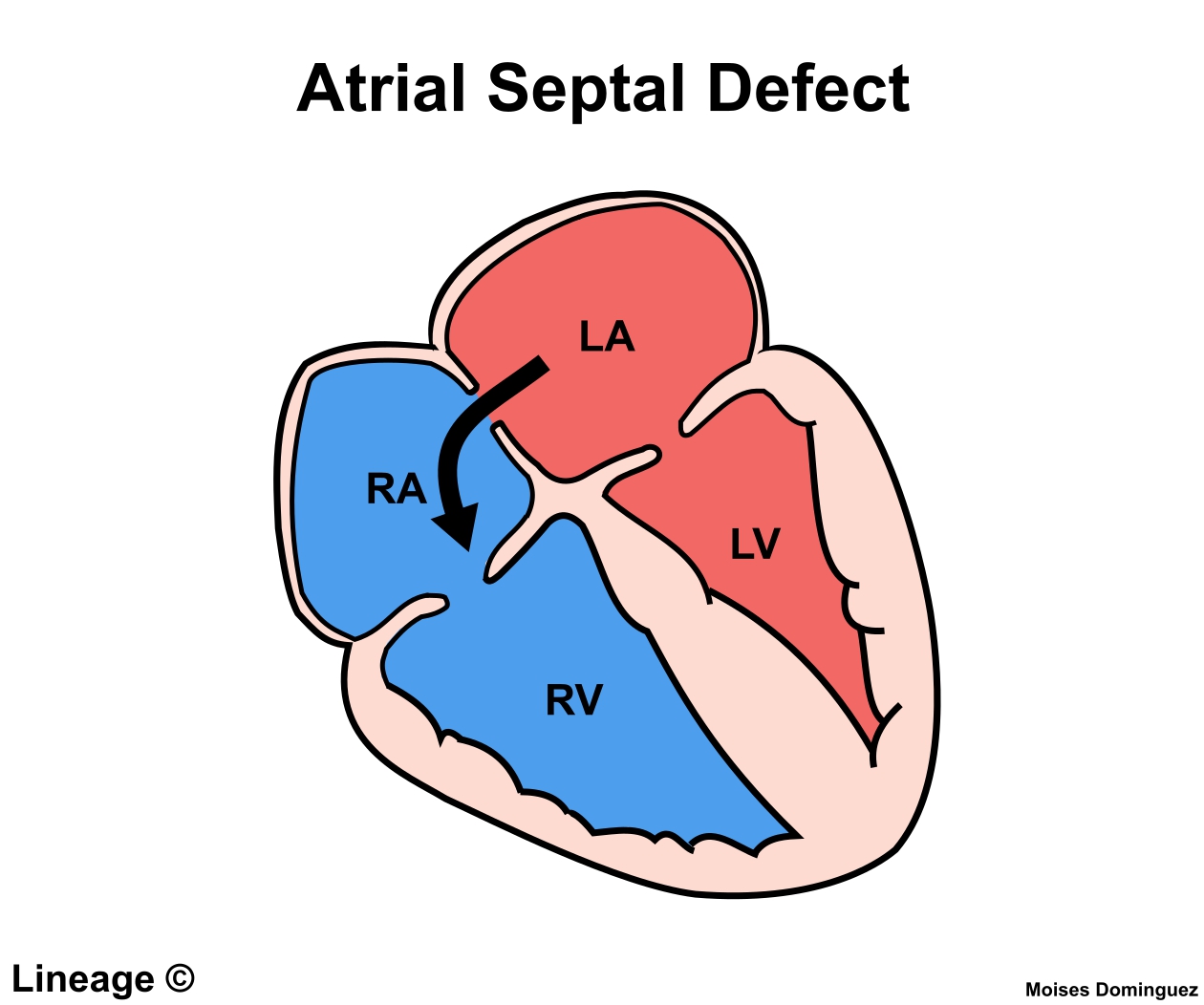
The primitive atrium is divided in two by joining of several structures. From the roof of the primitive atrium descends the septum primum, which grows towards the endocardial cushions within the atrial canal. Right before the septum primum fuses with the endocardial cushions there's a temporary space called the foramen primum. Once they fuse a new opening forms in the middle of the septum primum called the ostium secundum or foramen secundum. To the right of the septum primum and also coming down from the roof of the primitive atrium, descends a semilunar-shaped partition called the septum secundum. The free edges of the septum secundum produce an orifice called foramen ovale, which closes after birth when the septum primum and secundum fuse to each other completing the formation of the atrial septum.
The atrial canal is in turn divided into a right and left side by the atrioventricular septum, which originates from the union of the dorsal and ventral endocardial cushion. The right side of the atrial canal will become the tricuspid valve and the left will become the bicuspid valve.
Defects in producing the AV septum produces atrioventricular septal defects, including a persistent AV canal and tricuspid atresia.
Step 3(b): Ventricular septation[edit]
The floor at the midline of the primitive ventricle produces the interventricular septum, separating the chamber in two. The IV septum grows upward towards the endocardial cushion. As it grows, a foramen appears, the interventricular foramen, which later is closed by the non-muscular IV septum.
Defects in producing the IV septum causes ventricular septal defects, which communicate both ventricles.
Step 4: Outflow tract septation[edit]
The truncus arteriosus and the adjacent bulbus cordis partition by means of cells from the neural crest.[1] Once the cells from the truncal ridge meet with the cells from the bulbar ridge they twist around each other in a spiral orientation as they fuse and form the aorticopulmonary septum.[2] This will end dividing the aorta from the pulmonary trunk.[3]

Defects in this process is known as aortopulmonary septal defect, and causes persistent truncus arteriosus, unequal division of the truncus arteriosus, transposition of the great arteries, aortic and pulmonary valve stenosis or tetralogy of fallot.
Step 5: Heart valve formation[edit]
The heart valves are formed.
Defects in this process are known as valvular heart disease.
References[edit]
- ^Maschhoff KL, Baldwin HS (2000). 'Molecular determinants of neural crest migration'. Am. J. Med. Genet. 97 (4): 280–8. doi:10.1002/1096-8628(200024)97:4<280::AID-AJMG1278>3.0.CO;2-N. PMID11376439.
- ^Kirby ML, Gale TF, Stewart DE (1983). 'Neural crest cells contribute to normal aorticopulmonary septation'. Science. 220 (4061): 1059–61. doi:10.1126/science.6844926. PMID6844926.
- ^Jiang X, Rowitch DH, Soriano P, McMahon AP, Sucov HM (2000). 'Fate of the mammalian cardiac neural crest...'. Development. Cambridge, England. 127 (8): 1607–16. PMID10725237.
External links[edit]

- ]http://php.med.unsw.edu.au/embryology/index.php/Cardiac_Embryology Cardiac embryology] at UNSW
The USMLE Committee for Individualized Review (CIR) meets periodically throughout the year to review cases involving allegations of irregular behavior by applicants and/or examinees.
At its recent meetings, the CIR considered multiple cases involving the following:
- falsifying information, including the creation of falsified score reports
- seeking to obtain unauthorized access to examination materials
- communicating about specific test items, cases, and/or answers with other examinees
- providing unauthorized access to examination content on the internet
- applying for and/or attempting to take an examination when ineligible
- making notes on test day on something other than materials provided
- failure to follow test center instructions, including typing past the ‘End Patient Note’ announcement in Step 2 Clinical Skills
Actions taken by the CIR at its recent meetings included:
- annotating individual USMLE records with a finding of irregular behavior
- barring access to USMLE for periods up to 3 years
- reporting the finding of irregular behavior to the disciplinary data bank (Physician Data Center [PDC]) at the Federation of State Medical Boards (FSMB). State medical boards routinely query this data bank as part of their licensing processes
- canceling the examinee’s score because the validity of a passing level score is in question
Primum Step 3 Program Schedule
As evidenced by the sanctions listed above, a finding of irregular behavior carries significant potential impact. USMLE applicants and examinees are reminded to read the USMLE Bulletin of Information carefully, follow the rules of conduct during testing, and refrain from any pre- or post-examination conduct deemed to be irregular behavior.
Primum Step 3 Program Reviews
Applicants and examinees are also encouraged to watch the USMLE Security Video.
The USMLE is committed to maintaining the integrity of its examination so that state medical boards may continue to rely upon it as an integral part of their decision-making process for licensure. Applicants and examinees are advised to observe all USMLE policies and procedures to avoid the potentially significant implications arising from a finding of irregular behavior.
USMLE encourages you to provide information about cheating and other activity of which you are aware that may compromise the security and integrity of USMLE. Please use the contact form on the USMLE website to report such information.
ECFMG Policies and Procedures Regarding Irregular Behavior
ECFMG also regularly reviews allegations of irregular behavior in conjunction with its programs and services. ECFMG programs and services include but are not limited to, registering international medical students and graduates for USMLE Step 1, Step 2 CK, and Step 2 CS; ECFMG Certification; ERAS Support Services at ECFMG; the Exchange Visitor Sponsorship Program; the ECFMG International Credentials Services; and the Electronic Portfolio of International Credentials.
Primum Step 3 Program Manual
If the ECFMG Medical Education Credentials Committee determines that an individual engaged in irregular behavior, a permanent annotation to that effect will be included in the individual’s ECFMG record and will be provided to third parties in certain reports.
If it is determined that an individual engaged in irregular behavior, the individual also will be subject to serious sanctions. These sanctions may include, but are not limited to:
Primum Step 3 Program Download
- Barring the individual from future examinations;
- Barring the individual from ECFMG Certification and/or other ECFMG programs; and
- Revoking the individual’s Standard ECFMG Certificate.
Individuals who apply to ECFMG programs and services should be familiar with the ECFMG Policies and Procedures Regarding Irregular Behavior, which define irregular behavior and outline possible sanctions. Representative examples of allegations of irregular behavior and actions taken by the ECFMG Medical Education Credentials Committee are also available on the ECFMG website.